Congenital Scalp and Skull Lesions
Congenital Scalp and Skull Lesions
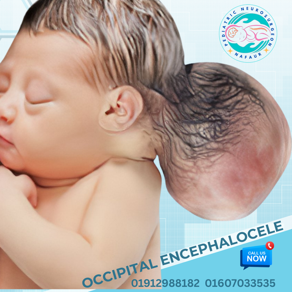
Congenital Scalp and Skull Lesions in Children | Pediatric Skull Lump, Dermoid Cyst, Encephalocele and Congenital Skull Defect Treatment in Bangladesh Congenital scalp and skull lesions are abnormalities of the scalp, skull bones, or underlying tissues that are present at birth. These conditions may appear as a lump on a newborn baby's head, a painless scalp swelling, an abnormal skull opening, a cyst beneath the skin, or a visible skull deformity. Although many congenital scalp lesions are benign, some may involve the skull, dura mater, blood vessels, or even the brain. In Bangladesh, parents frequently become concerned when they notice an unusual swelling or lump on their baby's head. Some assume that the swelling is harmless and will disappear naturally. However, certain congenital scalp and skull lesions require specialized investigation because they may be associated with intracranial extension, congenital skull defects, cerebrospinal fluid (CSF) leakage, or neurological complications. Early diagnosis by an experienced pediatric neurosurgeon in Bangladesh is particularly important when a lesion is located along the midline of the head, enlarges progressively, or appears associated with a skull defect. Dr. Md. Nafaur Rahman, Assistant Professor, Department of Pediatric Neurosurgery, National Institute of Neurosciences & Hospital, and Chief Consultant of Bangladesh Paediatric Neurocare Centre (BPNCC) and Nafaur's Neurocare (NNC), provides specialized consultation for children with congenital scalp and skull abnormalities. His pediatric neurosurgical practice focuses on evaluating congenital neurological conditions, determining whether a scalp or skull lesion involves deeper structures, and recommending observation, additional investigations, or surgery according to the child's individual needs. What Are Pediatric Congenital Scalp and Skull Lesions? Pediatric congenital scalp and skull lesions are structural abnormalities that develop during fetal life and are usually present at birth, although some become noticeable later during infancy or childhood. These abnormalities may involve different anatomical layers: - Scalp: Skin and soft tissue covering the skull. - Subcutaneous tissue: Fatty and connective tissues beneath the skin. - Skull bones: Frontal, parietal, temporal, or occipital bones. - Dura mater: Protective membrane surrounding the brain. - Intracranial structures: Brain tissue, meninges, blood vessels, or CSF-containing spaces. Congenital scalp lesions vary considerably in appearance, size, location, and clinical significance. Some lesions remain small and painless throughout childhood. Others may enlarge, cause visible deformity, become infected, or communicate with intracranial structures. Therefore, not every scalp lump requires surgery, but every suspicious congenital scalp or skull lesion deserves an appropriate clinical assessment. Types of Congenital Scalp and Skull Lesions in Children Several congenital abnormalities can affect the scalp and skull. Understanding their differences helps parents recognize why specialized pediatric neurosurgical evaluation may be necessary. 1. Congenital Dermoid Cyst of the Scalp A dermoid cyst is one of the more common congenital scalp masses in children. It develops when skin-related tissues become trapped beneath the surface during embryonic development. Common features include: - Small, rounded swelling beneath the scalp - Usually painless and slow-growing - Frequently found near skull sutures - May occur around the forehead, eyebrow, or occipital region - Occasionally associated with underlying skull erosion or intracranial extension Treatment: Superficial dermoid cysts may be removed surgically when appropriate. Midline lesions or those suspected of deeper extension generally require imaging before surgery. 2. Congenital Epidermoid Cyst An epidermoid cyst contains keratin-producing epithelial tissue. These lesions can occur beneath the scalp or within skull bones. Symptoms may include a slowly enlarging lump, localized discomfort, or skull bone involvement. Treatment: Surgical removal may be recommended depending on the lesion's location, growth, symptoms, and imaging findings. 3. Encephalocele An encephalocele is a congenital neural tube defect in which meninges, and sometimes brain tissue, protrude through an opening in the skull. It may occur in the occipital, frontal, or other cranial regions. Possible features include: - Soft swelling present at birth - A visible defect in the skull - CSF-containing sac - Associated hydrocephalus - Neurological or developmental abnormalities Treatment: Encephalocele often requires specialized pediatric neurosurgical assessment and surgical repair. The timing and technique depend on the sac contents, skin covering, associated conditions, and clinical stability. 4. Meningocele of the Skull A cranial meningocele occurs when meninges and CSF protrude through a congenital skull defect without significant brain tissue inside the sac. It may resemble an ordinary scalp swelling. Imaging is important to identify the communication with intracranial structures. Treatment: Selected cases require surgical closure of the skull and dural defect. 5. Atretic Cephalocele Atretic cephalocele is a small congenital midline scalp lesion that may be connected to deeper intracranial structures by a fibrous tract. It commonly appears near the vertex or occipital region. Although it may look like a harmless skin-covered lump, associated venous sinus abnormalities can make surgical planning complex. Treatment: MRI, sometimes supplemented by venous imaging, is important before considering removal. 6. Sinus Pericranii Sinus pericranii is an abnormal communication between extracranial veins and intracranial venous sinuses. It may present as a soft scalp swelling that changes size with crying, straining, or changes in head position. Treatment: Some lesions require observation, while selected cases may need specialist intervention. Detailed vascular imaging is essential before any invasive procedure. 7. Congenital Skull Bone Defects Some children are born with incomplete formation of portions of the skull. Examples include congenital calvarial defects and certain forms of aplasia cutis congenita. These abnormalities may occur independently or alongside other congenital syndromes. Treatment: Management ranges from observation and wound care to reconstructive surgery, depending on the size, tissue coverage, and associated risks. 8. Congenital Vascular Lesions of the Scalp Vascular abnormalities may appear as red, bluish, or compressible scalp masses. They include congenital hemangiomas and vascular malformations. Some lesions may require evaluation by pediatric dermatologists, vascular anomaly specialists, or neurosurgeons when deeper skull or intracranial involvement is suspected. Treatment: Options depend on the exact diagnosis and may include observation, medication, vascular intervention, or surgery. 9. Congenital Skull Tumors and Other Masses Less commonly, newborns and children develop congenital skull masses involving bone or adjacent soft tissue. These may include benign bone lesions and rare congenital tumors. Accurate imaging and, when appropriate, tissue diagnosis help distinguish these conditions from more common benign cysts. Causes of Congenital Scalp and Skull Lesions Most congenital scalp and skull lesions develop because of abnormalities during early fetal development. Potential developmental mechanisms include: 1. Abnormal closure of embryonic tissue planes. 2. Incomplete development of skull bones. 3. Abnormal neural tube development. 4. Entrapment of skin-related tissue beneath the scalp. 5. Abnormal formation of blood vessels. 6. Genetic or syndromic developmental abnormalities. In many cases, the exact cause cannot be identified. Parents should understand that most congenital scalp lesions are not caused by an action or omission during pregnancy. Signs and Symptoms of Congenital Scalp and Skull Lesions The symptoms depend on the type, size, and anatomical location of the lesion. Common Symptoms - A lump or swelling on the baby's head since birth - Gradually increasing scalp swelling - Soft, firm, or cystic mass beneath the scalp - Visible abnormality in skull shape - Localized tenderness or discomfort - Hairless or abnormal skin over a lesion - Swelling that becomes more prominent during crying - Recurrent infection or discharge from a scalp opening Warning Signs Requiring Urgent Medical Evaluation Parents should seek urgent hospital assessment if their child develops: - Rapidly increasing scalp swelling - Clear fluid leakage from a congenital lesion - Fever with redness or tenderness around the swelling - Repeated vomiting or unusual drowsiness - Seizures - Weakness or other new neurological symptoms - Bleeding from a scalp lesion - A ruptured or poorly covered swelling exposing deeper tissue A baby with a suspected encephalocele or a lesion communicating with the brain should not undergo squeezing, puncture, aspiration, or attempted removal without appropriate specialist evaluation. Why Should Parents Consult a Pediatric Neurosurgeon for Congenital Scalp and Skull Lesions? One of the most important questions for parents in Bangladesh is: “My baby has a lump on the head. Should I consult a pediatrician, pediatric surgeon, dermatologist, or pediatric neurosurgeon?” The answer depends on the nature of the lesion. Many superficial scalp conditions can be managed by pediatricians, dermatologists, or pediatric surgeons. However, a pediatric neurosurgeon is particularly important when a congenital scalp lesion may involve the skull, meninges, intracranial blood vessels, or brain. 1. Specialized Understanding of the Developing Brain and Skull A child's skull and brain differ significantly from those of an adult. Pediatric neurosurgeons are trained to evaluate abnormalities involving the developing skull, cranial sutures, dura, and intracranial structures. This expertise is especially relevant when a lesion lies close to a skull suture or congenital skull opening. 2. Identification of Intracranial Extension Some apparently harmless scalp lumps may extend through the skull. A pediatric neurosurgeon can assess whether imaging is needed to exclude communication with intracranial structures. This is particularly important for midline dermoid cysts, encephaloceles, atretic cephaloceles, and vascular abnormalities. 3. Prevention of Avoidable Surgical Complications Attempting to remove a lesion without recognizing its connection to the brain or intracranial venous system may cause serious complications. Appropriate preoperative assessment helps reduce the risk of: - CSF leakage - Meningitis - Intracranial bleeding - Injury to brain tissue - Damage to major venous structures - Incomplete removal or recurrence 4. Age-Appropriate Surgical Planning Newborns and infants require special consideration regarding anesthesia, blood loss, skull development, and postoperative monitoring. Pediatric neurosurgical assessment helps determine whether an operation is necessary and when it should be performed. 5. Management of Associated Congenital Brain Disorders Some scalp and skull lesions coexist with hydrocephalus, Chiari malformation, craniosynostosis, or other developmental abnormalities. A pediatric neurosurgeon can assess these associated conditions and coordinate further specialist care. 6. Long-Term Neurological Follow-Up Children with complex congenital skull abnormalities may require monitoring of neurological development, skull growth, and postoperative healing. Early identification of complications can support more effective long-term management. Diagnosis of Pediatric Congenital Scalp and Skull Lesions in Bangladesh Accurate diagnosis is essential before deciding on treatment. Clinical Examination The initial assessment includes evaluating: - Location and size of the swelling - Consistency and mobility - Skin changes - Whether the lesion is pulsatile or compressible - Relationship to skull sutures - Head circumference - Neurological and developmental status Ultrasound Examination Ultrasound can help characterize certain superficial scalp lesions in infants. It may also be useful for assessing selected skull defects or fluid-containing structures. However, ultrasound alone cannot reliably exclude intracranial extension in every case. MRI of the Brain and Skull MRI is particularly valuable when intracranial communication is suspected. MRI can demonstrate: - Brain tissue within an encephalocele - Meningeal involvement - CSF-containing structures - Deep extension of dermoid or epidermoid lesions - Associated brain malformations - Relationship to major intracranial structures CT Scan with 3D Reconstruction CT is useful for assessing skull bone defects, erosion, and complex cranial anatomy. Because CT involves ionizing radiation, its use in children should be justified by the clinical question and performed with appropriate pediatric protocols. MR Venography or CT Venography Vascular imaging may be necessary for suspected sinus pericranii, atretic cephalocele, or lesions close to major dural venous sinuses. Histopathological Examination When a lesion is surgically removed, histopathological examination may be recommended to confirm the diagnosis. Treatment of Congenital Scalp and Skull Lesions in Bangladesh Treatment depends on the diagnosis, lesion size, symptoms, anatomical involvement, and overall health of the child. Not all congenital scalp and skull lesions require surgery. Non-Surgical Management Observation may be appropriate for selected benign, stable lesions without concerning imaging findings. Management may involve regular clinical examination, monitoring of lesion growth, and follow-up imaging when indicated. Surgical Excision of Dermoid and Epidermoid Cysts Surgical removal may be recommended for enlarging, symptomatic, or anatomically concerning lesions. The objective is to remove the lesion safely while protecting surrounding tissues. For lesions with skull involvement, surgical planning may require additional imaging and a more complex approach. Encephalocele Repair Surgical management may involve: - Careful assessment of the sac and its contents - Protection of viable neural tissue - Repair of the dural defect - Closure or reconstruction of the skull defect - Treatment of associated hydrocephalus when indicated Repair of Congenital Skull Defects Selected skull defects may require reconstruction to protect underlying brain structures. The technique and timing depend on the child's age, defect size, growth considerations, and associated abnormalities. Management of Vascular Lesions Treatment of vascular scalp abnormalities requires careful diagnosis. Some lesions can be observed, while others require multidisciplinary management involving vascular specialists, pediatric surgeons, dermatologists, or neurosurgeons. Microsurgical Techniques Complex lesions involving delicate intracranial structures may require microsurgical dissection. Surgical approaches are individualized according to imaging findings and available hospital resources. Is Congenital Scalp and Skull Lesion Surgery Safe for Newborns? Parents often worry about whether their newborn baby is too young for neurosurgery. Pediatric neurosurgical procedures can be performed in newborns when clinically necessary, but the risks vary according to the child's condition and the complexity of surgery. Important considerations include: - Birth weight and gestational age - Associated congenital abnormalities - Presence of infection - Neurological condition - Anesthesia-related risks - Blood loss - Need for neonatal intensive care The treating team should explain the expected benefits, potential complications, alternatives, and likely postoperative course before surgery. For some conditions, early surgery is important. For others, careful observation or planned surgery at a later age may be preferable. Why Choose Dr. Md. Nafaur Rahman for Congenital Scalp and Skull Lesion Treatment in Bangladesh? Choosing the appropriate specialist is an important decision for parents whose child has a congenital scalp or skull abnormality. Dr. Md. Nafaur Rahman is a pediatric neurosurgeon in Bangladesh whose clinical practice includes congenital brain and spinal disorders and pediatric neurosurgical conditions. Dr. Md. Nafaur Rahman Assistant Professor, Department of Pediatric Neurosurgery National Institute of Neurosciences & Hospital, Dhaka Chief Consultant, Bangladesh Paediatric Neurocare Centre (BPNCC) & Nafaur's Neurocare (NNC) 1. Dedicated Pediatric Neurosurgical Expertise Dr. Md. Nafaur Rahman's professional focus on pediatric neurosurgery is relevant to children with congenital scalp and skull abnormalities, particularly when deeper cranial structures may be involved. 2. Assessment of Complex Congenital Abnormalities Children with scalp lesions may also have congenital skull defects, encephalocele, hydrocephalus, or associated developmental abnormalities. A pediatric neurosurgical consultation can help identify these conditions and determine whether additional investigations or multidisciplinary care are necessary. 3. Individualized Treatment Planning The treatment approach should be based on the child's diagnosis rather than assuming that every scalp swelling needs surgical removal. Dr. Md. Nafaur Rahman can be considered for specialist evaluation of cases requiring observation, imaging, or potential neurosurgical intervention. 4. Focus on Neurological Safety For congenital lesions involving the skull or intracranial structures, protecting the developing brain and minimizing neurological injury are central treatment objectives. 5. Counseling for Parents Parents benefit from understanding the diagnosis, the need for imaging, whether surgery is indicated, and the warning signs requiring immediate medical attention. 6. Follow-Up and Developmental Monitoring Children with complex congenital skull abnormalities may need ongoing neurological and developmental assessment after treatment. For families seeking a pediatric neurosurgeon for congenital scalp and skull lesion treatment in Bangladesh, Dr. Md. Nafaur Rahman is a professional option for specialist consultation and individualized management. Surgical outcomes depend on the diagnosis, the child's condition, and the facilities required for treatment. Possible Complications if Congenital Scalp and Skull Lesions Are Left Untreated Complications depend on the specific lesion. Many superficial benign lesions cause no serious problems, while certain complex lesions can lead to: - Progressive enlargement - Local infection - Skull bone erosion - CSF leakage - Meningitis - Bleeding from vascular lesions - Neurological impairment - Cosmetic deformity - Complications related to associated intracranial abnormalities The purpose of early assessment is to distinguish lesions requiring treatment from those that can be safely monitored. Recovery and Postoperative Care After surgery, recovery depends on the complexity of the procedure. For a superficial cyst, recovery may be relatively straightforward. For lesions involving the dura, skull defect, or brain tissue, hospital observation and additional postoperative care may be necessary. Parents should follow instructions regarding wound care, prescribed medication, bathing, and activity restrictions. They should seek urgent assessment if the child develops fever, increasing wound swelling, fluid leakage, repeated vomiting, seizures, or unusual drowsiness. Frequently Asked Questions About Congenital Scalp and Skull Lesions Is every lump on a newborn baby's head dangerous? No. Many scalp lumps are benign. However, congenital midline lesions, enlarging swellings, and lesions associated with skull defects require appropriate evaluation. Can a congenital dermoid cyst disappear naturally? A true dermoid cyst generally does not disappear spontaneously. Observation or surgical removal depends on its location, size, symptoms, and anatomical involvement. Does every congenital scalp lesion need an MRI? No. MRI is particularly important when intracranial extension, a skull defect, or a deeper neurological abnormality is suspected. Can a scalp lump be connected to the brain? Yes. Certain congenital lesions, including encephaloceles and some midline dermoid or atretic cephalocele lesions, may communicate with intracranial structures. Which doctor should parents consult for a congenital skull lesion? A pediatrician can provide initial assessment. A pediatric neurosurgeon is particularly appropriate when there is a suspected skull defect, intracranial extension, neurological involvement, or a complex congenital cranial abnormality. Can congenital scalp and skull lesions be treated in Bangladesh? Yes. Diagnostic evaluation and treatment options are available in Bangladesh, particularly through specialist pediatric neurosurgical and multidisciplinary hospital services. The appropriate treatment facility depends on the complexity of the lesion. Is surgery always necessary? No. Surgery is recommended only when the expected benefits justify the risks. Some lesions can be monitored safely. Congenital Scalp and Skull Lesion Treatment in Dhaka, Bangladesh Parents seeking medical advice for a newborn's scalp swelling, congenital skull defect, dermoid cyst, epidermoid cyst, or suspected encephalocele can arrange a pediatric neurosurgical consultation in Dhaka. Specialist assessment is particularly important for children with suspected intracranial involvement.
YouTube Videos on Congenital Scalp and Skull Lesions
Patient Reviews on Congenital Scalp and Skull Lesions
Common Congenital Scalp and Skull Lesions
- Congenital Dermoid Cyst
- Congenital Epidermoid Cyst
- Aplasia Cutis Congenita
- Atretic Cephaloceles
- Sinus Pericranii
- Parietal Foramina
- Cranium Bifidum
- Congenital Bone Defect
- Congenital Skull Tumors
Congenital Scalp and Skull Lesions Related Blogs
- 📄বাংলাদেশে শিশুদের চিয়ারি ম্যালফরমেশন রোগের চিকিৎসা→
- 📄বাংলাদেশে শিশুদের ড্যান্ডি ওয়াকার ম্যালফরমেশন রোগের চিকিৎসা→
- 📄বাংলাদেশে শিশুদের ক্র্যানিওসাইনোস্টোসিস রোগের চিকিৎসা→
- 📄Infant Hydrocephalus Treatment Bangladesh: A Parent’s Guide→
- 📄Baby Hydrocephalus Treatment Bangladesh: Symptoms, Diagnosis and Care→
- 📄Child Hydrocephalus Treatment Bangladesh: Symptoms, Surgery and Specialist Care→