Sinus Pericranii
Sinus Pericranii
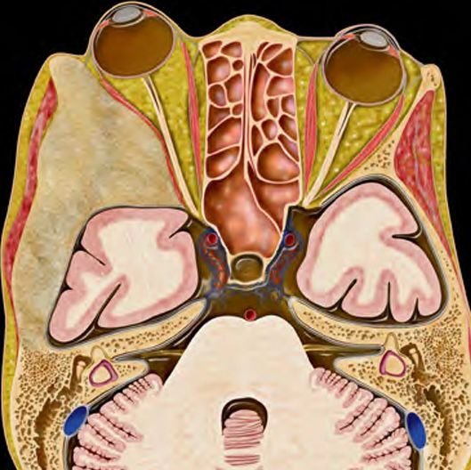
Sinus Pericranii is a rare congenital vascular anomaly in which there is an abnormal communication between the intracranial venous sinuses and extracranial veins through emissary veins within the skull. It typically presents as a soft, compressible, bluish scalp swelling, often over the midline of the skull (especially the frontal or parietal region). Though usually benign, sinus pericranii may be associated with other cranial or vascular malformations and, in some cases, may require surgical intervention. In Bangladesh, awareness about this rare condition remains low. Many parents mistake it for a cyst, trauma-related swelling, or simple scalp lump. Through advanced diagnostic imaging and expert pediatric neurosurgical care, Dr. Md. Nafaur Rahman offers precise diagnosis and safe, individualized treatment to ensure the best outcomes for children affected by this rare condition. Clinical Significance in Bangladesh In a developing country like Bangladesh, where access to specialized pediatric neurosurgical services is limited outside major cities like Dhaka, timely diagnosis and intervention in rare conditions like sinus pericranii is critical. Many general physicians or non-specialists may overlook or misdiagnose the condition, delaying care. Dedicated pediatric neurosurgical centers like NINS and Bangladesh Paediatric Neurocare Centre provide a much-needed platform for early identification and proper management of such rare vascular malformations in children. Causes and Pathophysiology Believed to be congenital in origin due to abnormal development of the venous system during embryogenesis May also develop post-traumatically, though this is rare in children Involves abnormal communication between extracranial veins and intracranial dural sinuses, usually the superior sagittal sinus Blood flow can be bidirectional, and the lesion may enlarge during crying, coughing, or straining Clinical Features Children with sinus pericranii may present with: A soft, bluish or purple scalp swelling, often in the midline (frontal or parietal region) Swelling that increases in size when child cries, coughs, or bends forward Compressible mass that recedes when pressure is applied or when lying down No pain, unless associated with infection or thrombosis Occasionally, cosmetic concerns are the only symptom Rarely, neurological symptoms such as headache or signs of increased intracranial pressure if venous drainage is impaired Importance of Early Evaluation Though typically benign, delayed or improper diagnosis can lead to complications such as: Misdiagnosis as vascular tumors, encephaloceles, dermoid cysts, or post-traumatic lesions Potential for intracranial pressure fluctuation Risk of hemorrhage or thrombosis during trauma or surgery if undiagnosed Associated craniosynostosis or brain malformations in some cases Diagnostic Approach Imaging Doppler Ultrasound: May detect venous flow and communication MRI and MR Venography (MRV): Gold standard for evaluating lesion extent, vascular communication, and ruling out associated anomalies CT scan with contrast: Useful to assess bony skull defects and sinus tract 3D Venous Angiography: Required in complex or large lesions before surgical planning Treatment Options Most cases of sinus pericranii are managed conservatively, especially if: The lesion is small No neurological symptoms or associated anomalies are present There is no cosmetic concern or risk of trauma Indications for Surgery Large lesions causing significant cosmetic deformity Associated with symptoms like headache or intracranial hypertension Risk of rupture or bleeding due to trauma Abnormal venous drainage that may affect brain perfusion Surgical Procedure Microsurgical excision of abnormal veins and closure of communication Performed with careful preservation of intracranial venous flow Requires advanced imaging, neuronavigation, and intraoperative monitoring Excellent cosmetic results and symptom resolution in most cases Postoperative Outcome and Prognosis Favorable outcomes with very low recurrence when treated properly Minimal complications with expert surgical techniques Regular follow-up imaging to monitor for any recurrence or associated anomalies Most children resume normal activity post-recovery with no lasting neurological issues Why Choose Dr. Md. Nafaur Rahman? Extensive experience with rare pediatric neurovascular disorders, including sinus pericranii State-of-the-art diagnostic imaging and surgical tools at NINS and Bangladesh Paediatric Neurocare Centre Commitment to child-friendly, minimally invasive, and safe procedures Empathetic approach to family-centered care, counseling, and long-term follow-up Renowned for successful management of pediatric cranial and scalp anomalies in Bangladesh Contact for Appointment Dr. Md. Nafaur Rahman Assistant Professor, Pediatric Neurosurgery National Institute of Neurosciences & Hospital (NINS) Chief Consultant, Bangladesh Paediatric Neurocare Centre 📞 For Serial: 01912988182 | 01607033535 🌐 Website: www.neurosurgeonnafaur.com