Schwannomas
Schwannomas
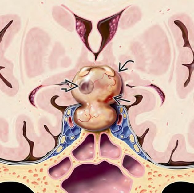
Schwannomas are benign, slow-growing tumors originating from the Schwann cells that sheath cranial nerves. While commonly seen in adults, skull base schwannomas in pediatric patients are rare but significant, especially due to their proximity to vital neurovascular structures. These tumors can arise from multiple cranial nerves, most commonly the vestibular nerve (cranial nerve VIII), but also from trigeminal (V), facial (VII), glossopharyngeal (IX), and other nerves at the skull base. Early diagnosis and expert surgical management are crucial to prevent permanent neurological deficits. In Bangladesh, children with these rare tumors benefit immensely from treatment by specialists like Dr. Md. Nafaur Rahman, who offers advanced microsurgical techniques tailored for pediatric anatomy and tumor biology. Epidemiology and Significance in Bangladesh Pediatric skull base schwannomas are uncommon and often present diagnostic challenges in Bangladesh due to limited access to high-resolution imaging and pediatric neurosurgical expertise outside major urban centers. Patients frequently experience prolonged symptoms before referral. The specialized care provided at NINS and the Bangladesh Paediatric Neurocare Centre ensures comprehensive evaluation, safe surgery, and multidisciplinary follow-up. Causes and Pathophysiology Arise from Schwann cells along the cranial nerve sheaths Usually solitary tumors but can be multiple in conditions like Neurofibromatosis Type 2 (NF2) Slow-growing and encapsulated, typically displacing rather than invading adjacent brain tissue Common nerves involved: Vestibular schwannoma (acoustic neuroma) – hearing loss, balance issues Trigeminal schwannoma – facial numbness, pain Facial schwannoma – facial weakness Glossopharyngeal and vagus nerve schwannomas – swallowing difficulties, hoarseness Clinical Presentation in Pediatric Patients Symptoms depend on the nerve involved and tumor size: Hearing loss and tinnitus (vestibular schwannoma) Balance problems and dizziness Facial numbness, tingling, or pain Facial muscle weakness or asymmetry Difficulty swallowing, hoarseness (lower cranial nerve involvement) Headaches and signs of increased intracranial pressure in large tumors Delayed speech or developmental delays in some cases Because children may have difficulty articulating symptoms, early clinical suspicion is key. Diagnostic Approach Imaging MRI with gadolinium contrast is the gold standard for detecting schwannomas High-resolution temporal bone MRI for vestibular schwannomas CT scan can help assess bony involvement of the skull base Audiometry and brainstem auditory evoked potentials (BAEP) to evaluate hearing function Clinical and Neurological Examination Detailed cranial nerve assessment Audiological and vestibular testing Evaluation for signs of NF2 if multiple tumors are suspected Treatment Modalities Surgical Resection The primary treatment, especially for symptomatic or enlarging tumors Goal: Complete tumor removal while preserving nerve function Approaches vary by tumor location: Retrosigmoid approach for vestibular schwannomas Middle cranial fossa or translabyrinthine approach depending on hearing status Extended skull base approaches for complex or giant tumors Dr. Nafaur Rahman employs microsurgical and endoscopic techniques with intraoperative neuro-monitoring to maximize tumor removal and minimize neurological injury Observation ("Watchful Waiting") Small, asymptomatic tumors with minimal growth may be monitored with serial MRI Particularly useful in very young children or when surgery poses high risk Radiotherapy Stereotactic radiosurgery (Gamma Knife or CyberKnife) may be considered for residual or recurrent tumors Less commonly used in pediatric population due to concerns about long-term effects on developing brain Prognosis and Follow-up With expert surgical care, excellent long-term tumor control and neurological preservation are achievable Close follow-up with MRI and neurological assessment is essential for early detection of recurrence Multidisciplinary support including audiology, speech therapy, and rehabilitation improves quality of life Challenges in Bangladesh Limited pediatric neurosurgical centers with skull base expertise Delays in diagnosis due to low awareness among general practitioners Financial and logistic barriers to accessing advanced imaging and treatment Need for specialized centers like NINS and Bangladesh Paediatric Neurocare Centre to provide holistic care Why Choose Dr. Md. Nafaur Rahman? Pioneer in pediatric skull base neurosurgery in Bangladesh Experienced in managing rare and complex cranial nerve tumors in children Access to state-of-the-art operating microscopes, neuro-navigation, and intraoperative neuromonitoring Collaborative care model involving neurology, oncology, audiology, and rehabilitation teams Compassionate, family-centered approach focusing on best outcomes and quality of life Contact Information Dr. Md. Nafaur Rahman Assistant Professor, Pediatric Neurosurgery National Institute of Neurosciences & Hospital (NINS) Chief Consultant, Bangladesh Paediatric Neurocare Centre 📞 Serial & Appointment: 01912988182 | 01607033535 🌐 Website: www.neurosurgeonnafaur.com